Digitized
podoscopy
Clinical use. It is used in pathologies of the developmental and
adult age of the foot especially if accompanied by areas of considerable dyskeratosis (calluses, tilomas).
Protocol. The patient is positioned barefoot in a static standing
position on the scanner for 5 seconds. Following the photograph of the sole of
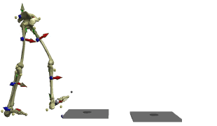
the foot is digitized. The software processes its image and through algorithms
allows you to correlate the topographical indicators with the underlying
proportional anatomical reconstruction of the foot bones. In this way it is
possible to relate the areas of hyperpressure with
the underlying anatomical structure affected by the pathology.
Procedures. The indicators used are structural of the foot:
breech index, metatarsal, mesopodal, isthmus, heel,
heel, heel half-circumference, skeletal analysis.
Execution times for the exam: 5 seconds.
Conclusions. Within 30 minutes the operator analyzes the data
(indicators), prints the graphs and concludes, delivering everything to the
patient in a sealed envelope.
The report written and signed by the doctor with the
diagnostic conclusions, prescriptions or therapeutic or clinical in-depth
recommendations (
Instrumental exams can then be repeated for normal
follow-up based on the indicators to be checked over time. If, in order to
check over time, the same doctor prescribes the tests to be repeated on a
company request