Three-dimensional
reconstruction of the spine
Clinical use. Scoliosis, paramorphisms,
dorsal and lumbar hyperhypociphosis are studied.
Protocol. The subject is invited to undress. It is prepared with
the positioning of adhesive skin markers on pre-established points in the
literature on the trunk, column, pelvis, upper limbs and is then invited to
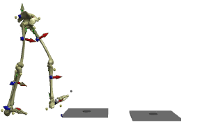
position itself on a platform at a distance preset by the cameras.
In a static standing position, the shots are taken in:
front, rear, right side and left side.
Procedures. The indicators that are studied for the deviations of
the column on the frontal and sagittal plane are:
1. Cobb degree measurement
2. angles of the cervico-dorsal
and dorso-lumbar curves
3. Others: linear distances between various landmarks
Execution time: 1 minute at acquisition if the patient collaborates
(moves little) and at each new acquisition if you want to test the variation of
the Cobb degrees and the cervico-dorsal, dorso-lumbar angles using simultaneously the calibrated stabilometry (see before) or various elevations under the
limbs.
Conclusions. Within 30 minutes the operator analyzes the data
(indicators), prints the graphs and concludes, delivering everything to the
patient in a sealed envelope.
The report written and signed by the doctor with the
diagnostic conclusions, prescriptions or therapeutic or clinical in-depth
recommendations (
Instrumental exams can then be repeated for normal
follow-up based on the indicators to be checked over time. If, in order to
check over time, the same doctor prescribes the tests to be repeated on a
company request.